
A painful Achilles tendon; first impressions and an ultrasound examination. Do the findings impact the rehabilitation?
A 61-year-old man presented with a painful Achilles region after slipping down a hill.
Ultrasound examination shows a markedly thickened Achilles tendon, heterogeneous, with microcystic change and collagen fascicular discontinuity, moderately increased vascularity, irregular inferior margin, thickened paratenon, and pain on pressure. There is no gap or fat pad infiltration.
First impressions show a tendon most closely fitting with Cook and colleagues’ definition of degenerative tendinopathy, but why then the pain?
One explanation is that there is acute on chronic tendinopathy (i.e., portions of healthy tissue within the tendon that have become degenerative).
Another explanation is that there is an acute tear, which is what, in fact, we saw.
This must surely impact rehabilitation. During the process of rebuilding tendon capacity, how does an acute tear change the way in which a tendon is loaded? Could the usual process of load progression for tendinopathy rehabilitation be detrimental to the healing of a tear?
Let us know what you think.
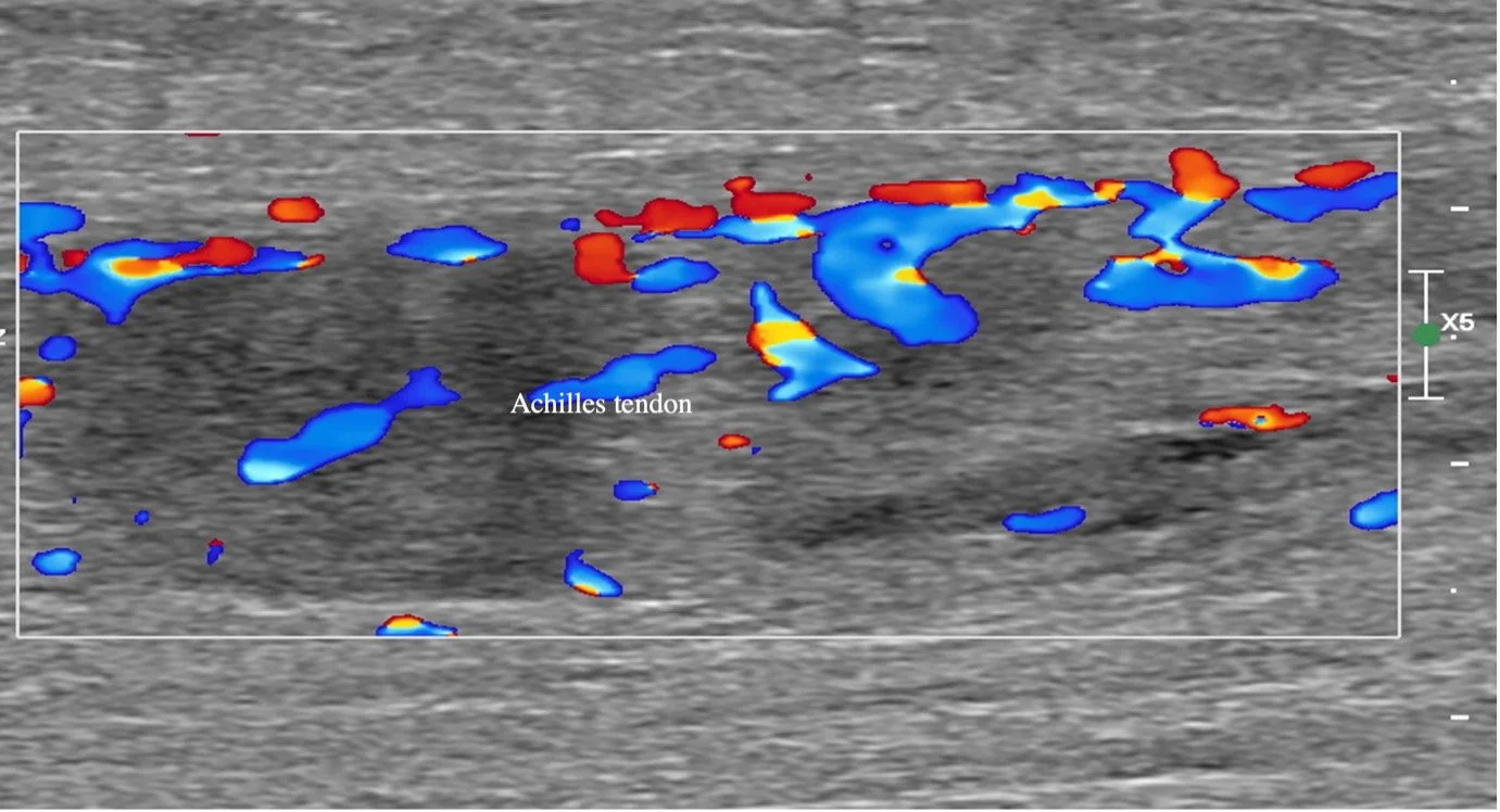
Image 1 Achilles tendon mid part in long view showing a markedly thickened, heterogeneous tendon with moderately increased vascularity.

Image 2 Achilles tendon mid-to-superior part in long view showing a markedly thickened, heterogeneous tendon with an ill-defined acute tear indicated by hashmarks and arrow.

Image 3 Achilles tendon mid-to-superior part in axial view showing a markedly thickened, heterogeneous tendon with an ill-defined acute tear indicated by hashmarks and arrow.