
Case studies and articles
The Sound Experience team publish regular educational case studies, research digests and opinions.

Corticosteroid injections in tendinopathy: why we will continue to follow guidelines.
From time to time we have a request for an injection into a tendon. With a clear rationale and usually a documented history of reasonable rehabilitation failing we may make an exception.
This post summarises a recently published article, incorporating a brief quality appraisal of recent evidence, and presents a coherent opinion reinforcing why we will continue to recommend against most tendon steroid injections.

A sit-down with Hamza & Naythan, the Stoddard Road Physios. Our technical ability (1/3).
A sit-down with Hamza & Naythan, the Stoddard Road Physios. Learning about our referrers, exploring themes on technical ability, the ultrasound report, and patient care.
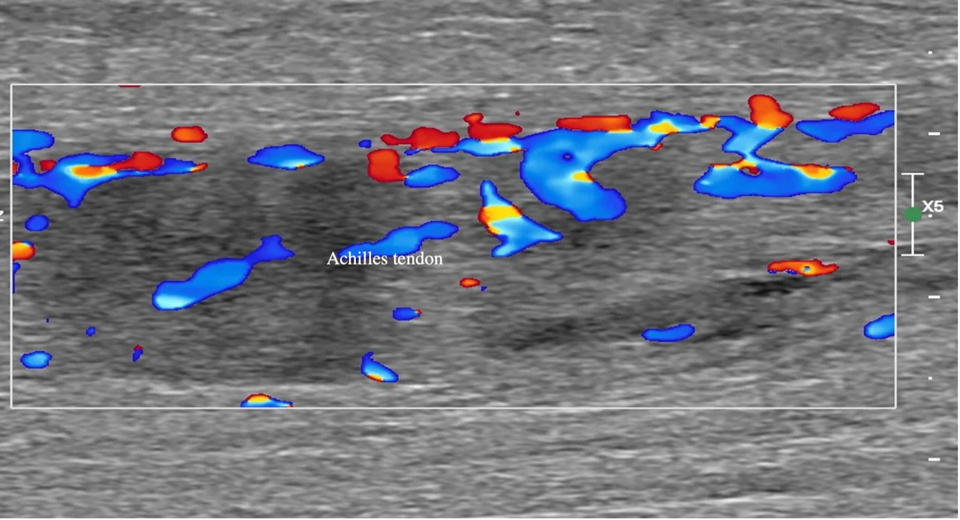
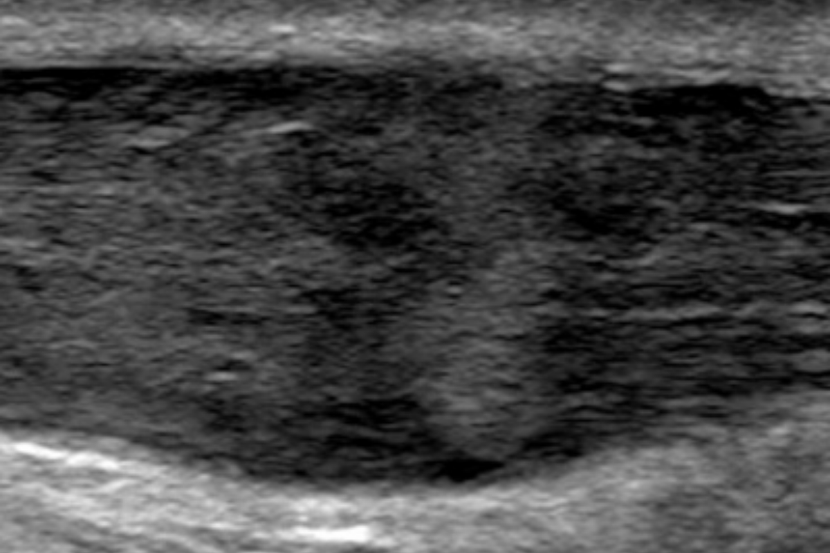
A painful Achilles tendon; first impressions and an ultrasound examination. Do the findings impact the rehabilitation?
After slipping down a hill, a 61-year-old man presented with a painful Achilles region. First impressions show a tendon most closely fitting with Cook and colleagues’ definition of degenerative tendinopathy, but why then the pain?
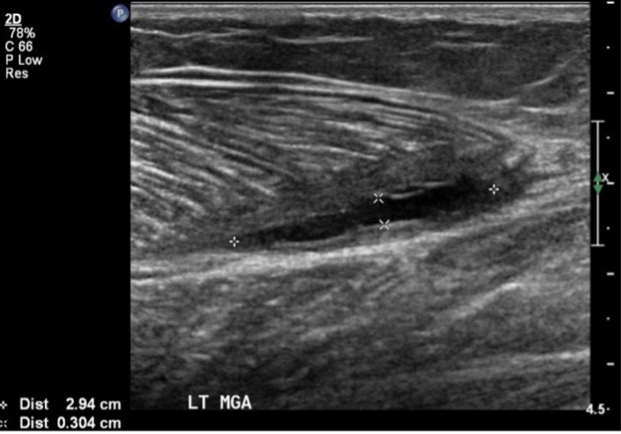
Muscle tears, grading muscle injury, and the ultrasound report. What’s new?
Muscle tears, Grading Muscle Injury and Reporting. What’s new? At our recent referrer’s evening, we spent time discussing our current approach to reporting and monitoring muscle tears.
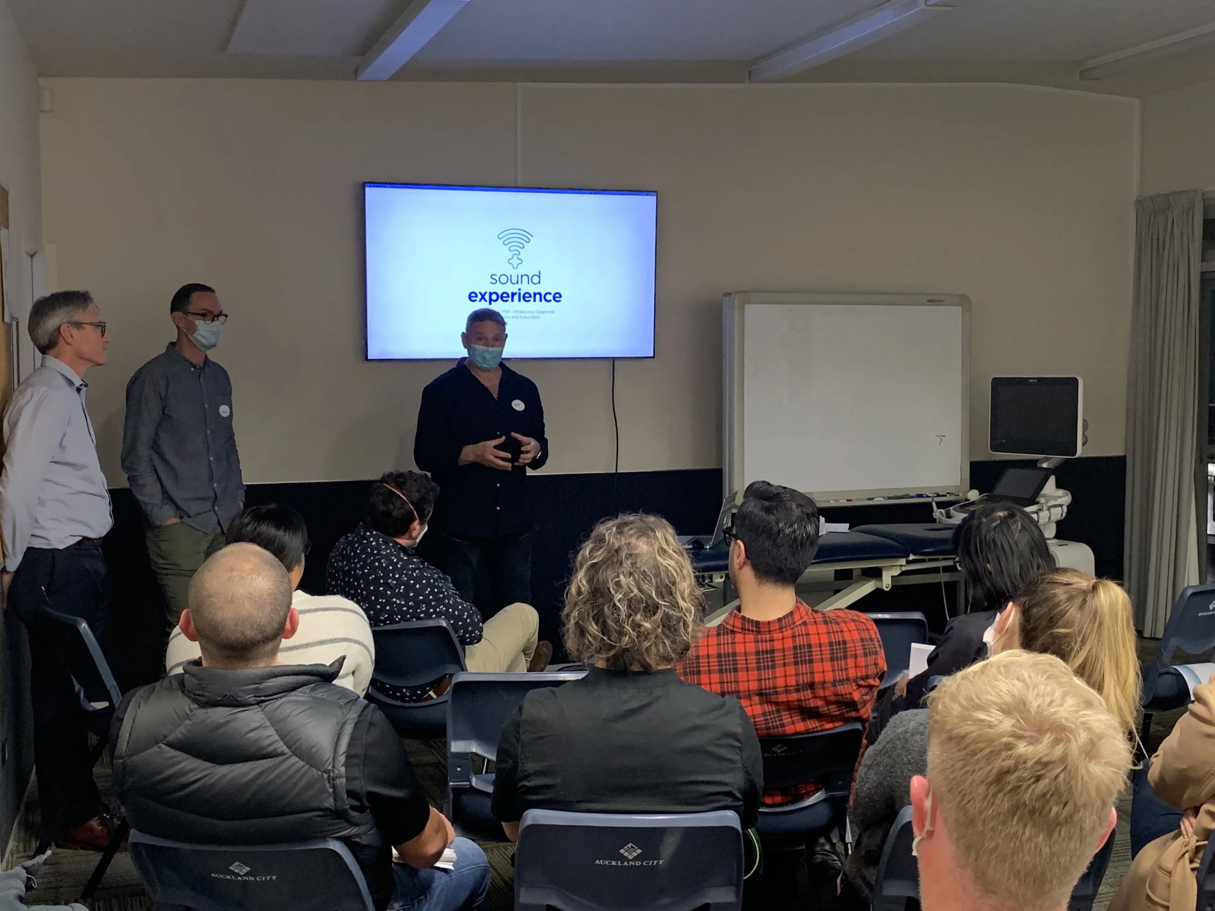
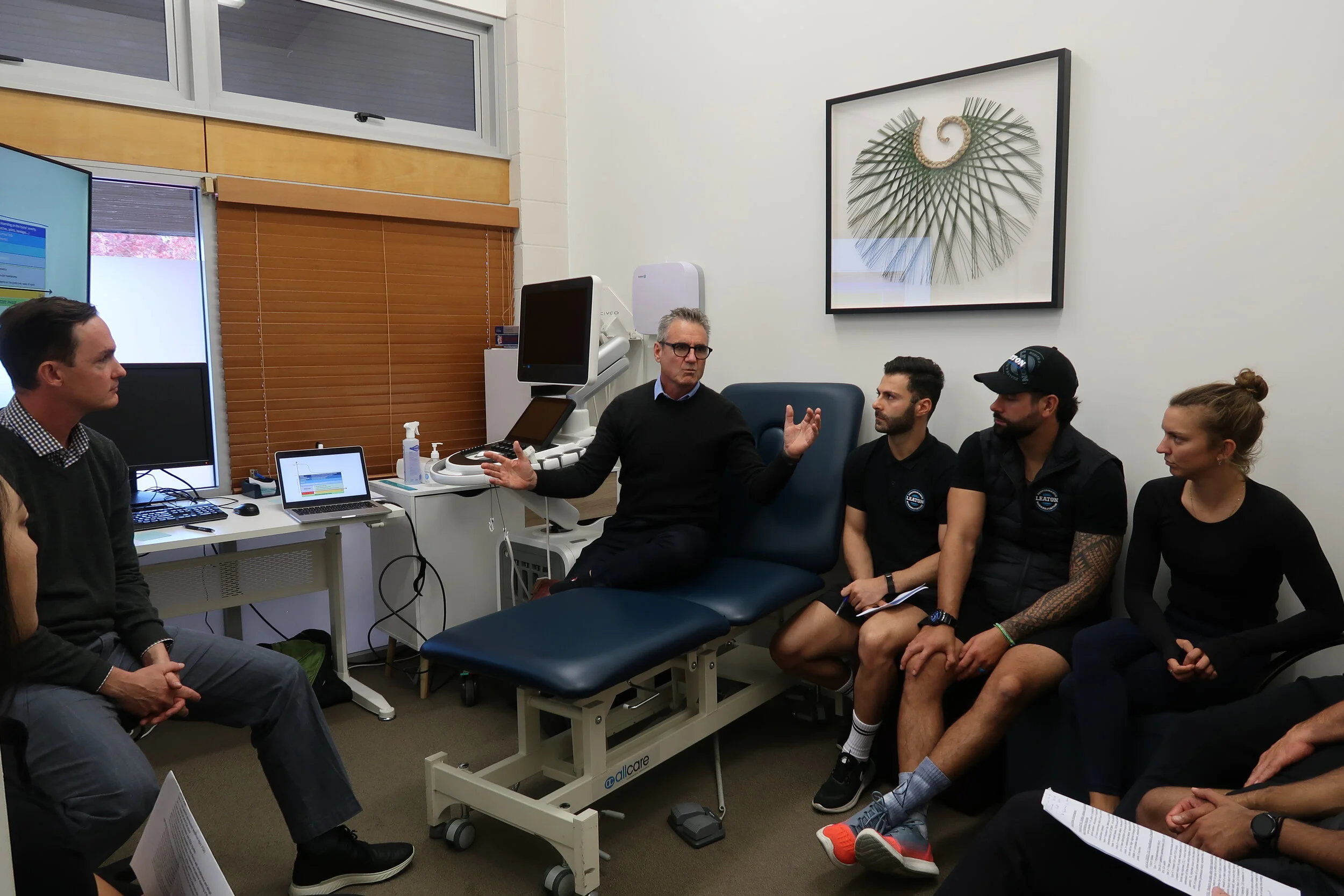
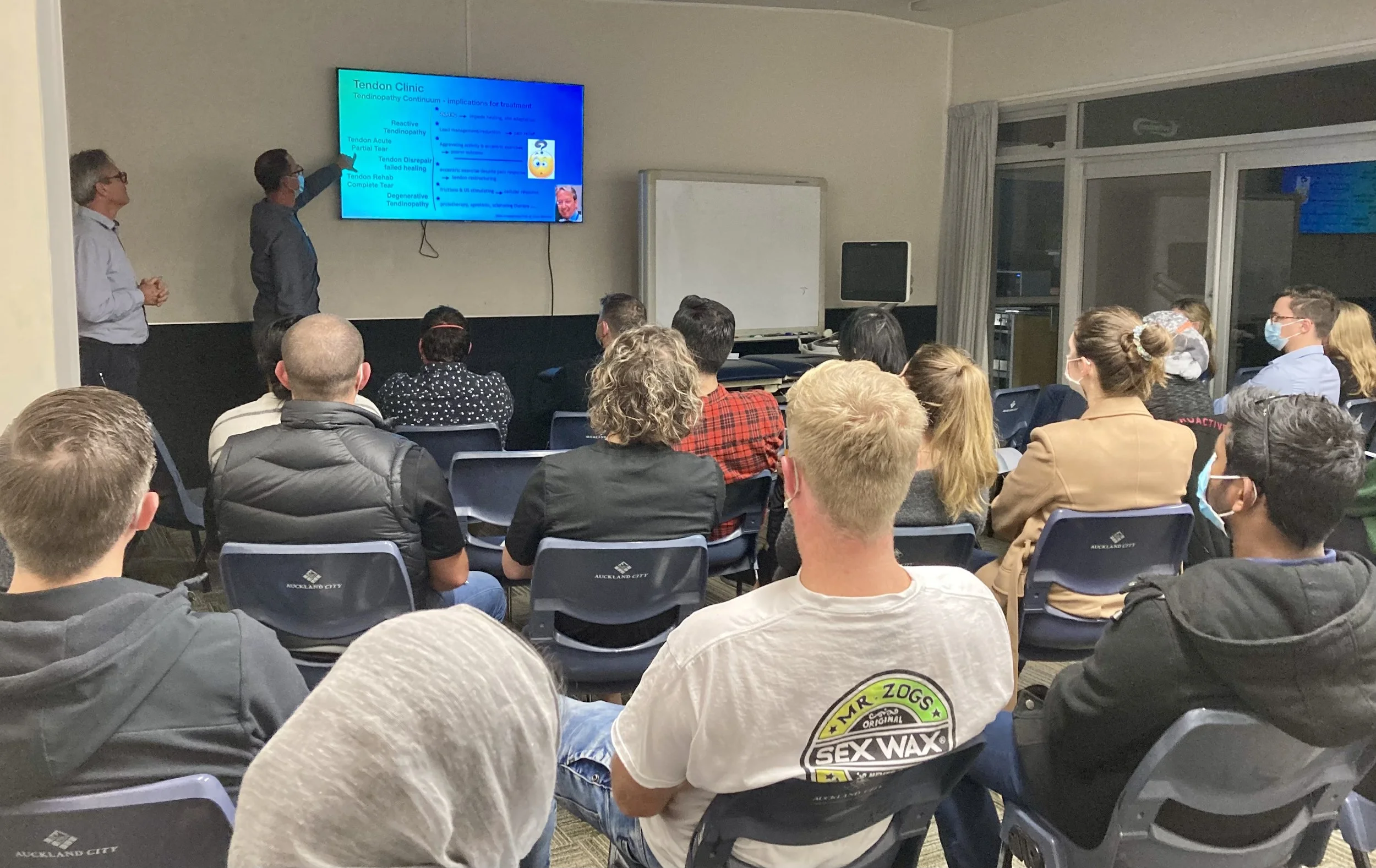
Musculoskeletal Ultrasound Information event – 29 Aug 2022
Musculoskeletal Ultrasound Information event – 29 Aug 2022. After a prolonged COVID interruption, we hosted a learning event, the first since July 2021.

Not Sever's Disease
“Not Sever’s Disease” A 10-year-old boy, very active, presents with a 4-month history of heel pain, aggravated by activity.

Our grading system is based on anatomy, not numbers.
Serial ultrasound examination of a quadriceps muscle injury. Our grading system is based on anatomy, not numbers. A 61-year-old man presented suffering from right anterior thigh pain following tripping and essentially doing the splits.

The “Quality Triangle”
The “Quality Triangle” describes the theory that within a project, there are three axes- cost, quality, and time- of which one is usually fixed, while the other two have an inverse relationship.
Annoying as that is, being conscious of the trade-offs and allowing for them or even actually planning for them, might be a sensible idea…
What’s in a name?
Sound Experience – is an unusual name for an ultrasound imaging business. People are curious about our business name and often ask what it means. Why the unconventional name for a conventional healthcare service?
Where do our muscle tear scanning intervals come from? (AKA our promise)
Scott Allen recently talked about our current thinking (yes, it may well change) regarding muscle tears. He mentioned three key points. We thought it might be sensible to add a little insight into how we arrived at the intervals and ultrasound tear evaluation.
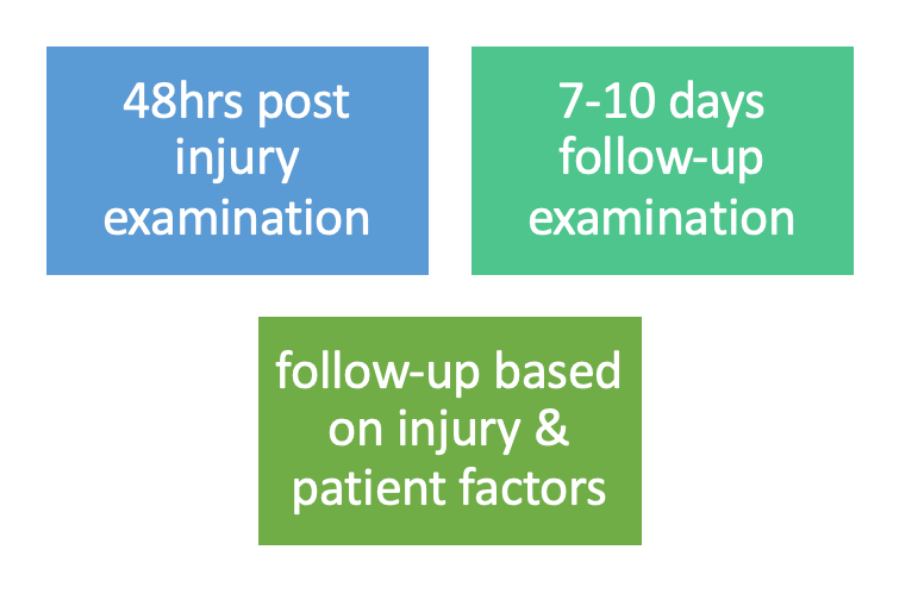
Let’s talk about ultrasound imaging in acute muscle injury
We had a small zoom meeting recently to start a conversation about muscle injury. This was an opportunity to share with referrers and other interested people our current interpretation of what the literature is telling us regarding the role of ultrasound imaging in the diagnosis and rehabilitation of acute muscle tears. There were three initial focus points we wanted to share:
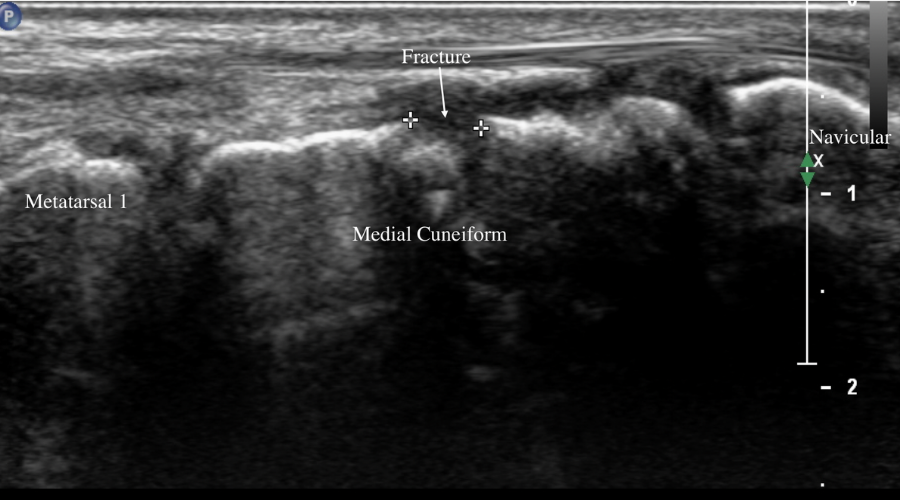
Foot injury: medial cuneiform fracture.
An 18-year-old woman presented suffering from left foot pain after having her left foot stood on while playing rugby at an out of town tournament three weeks prior. Following an ultrasound examination, x-ray and a finally a CT scan she was booked for surgery.
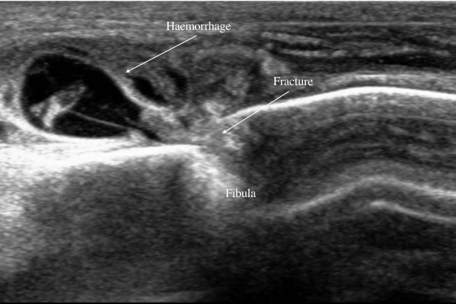
Ankle injury: spiral fibula fracture
A 60-year-old man presented with left ankle pain after rolling his left ankle. His ankle was extensively bruised and swollen. We explored the injury with ultrasound and X-ray and diagnosed spiral fibula fracture. Urgent surgery was required.
Finding fractures with ultrasound. Part 2
In part one, we left you with, why does detecting a fracture with ultrasound make a difference? The treatment pathway for fracture is different from that of soft tissue injury.
Finding fractures with ultrasound. Part 1
I am often asked, “Can ultrasound detect fracture?” The short answer, “Yes.” In this two part post, find out why this matters.
Indicators of intra-articular pathology on ultrasound. Part 2
In the second part of this two part post, learn why indicators of intra-articular pathology seen on ultrasound may require further investigation.
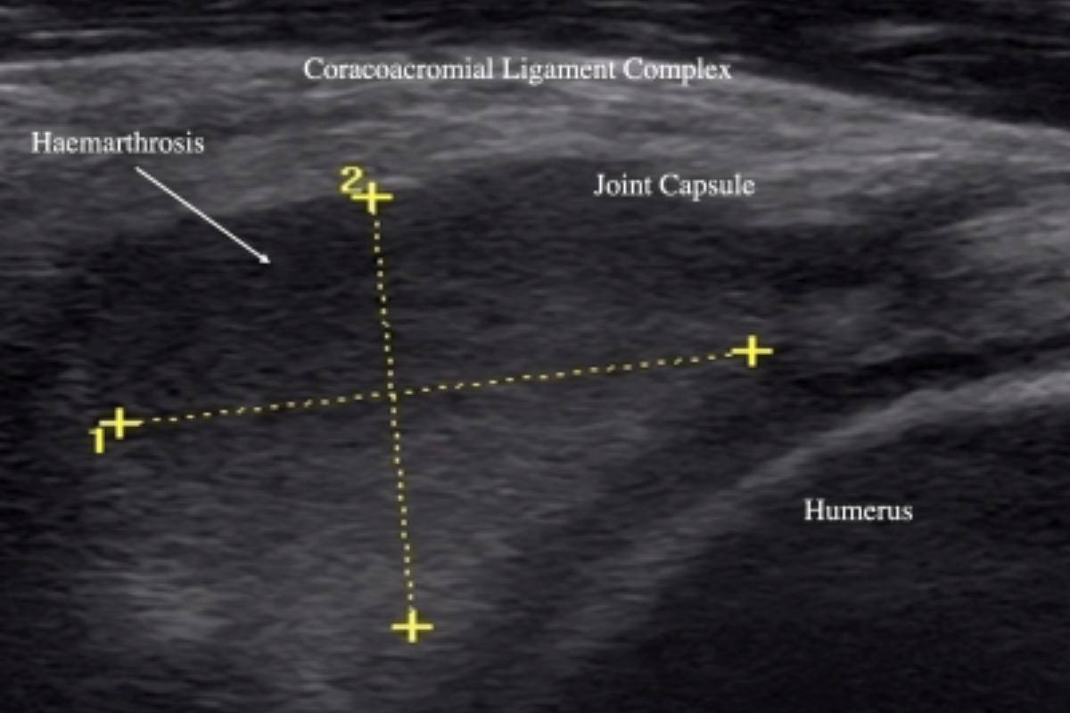
Indicators of intra-articular pathology on ultrasound. Part 1
In the first part of this two part post, learn why indicators of intra-articular pathology seen on ultrasound may require further investigation.